Main Page: Difference between revisions
No edit summary |
|||
| (91 intermediate revisions by 2 users not shown) | |||
| Line 1: | Line 1: | ||
== Our Facility == | === Our Facility=== | ||
MRI facility on the Faculty [http://www.uib.no/mofa/nyheter/2013/10/oppdrag-sjaa-det-skjulte website] | |||
<big>'''Bruker Pharmascan 70/16'''</big> | <big>'''Bruker Pharmascan 70/16'''</big> | ||
A 7.0 Tesla small animal magnetic resonance (MR) scanner was installed at the Vivarium, University of Bergen, in December 2004.<br> | A 7.0 Tesla small animal magnetic resonance (MR) scanner was installed at the Vivarium, University of Bergen, in December 2004 and was upgraded with new hardware in December 2012. <br> | ||
This MR scanner can be used for non-invasive diagnostic studies on small laboratory animals (rats, mice, small fish). | |||
Pathological | This MR scanner can be used for non-invasive diagnostic studies on small laboratory animals (rats, mice, small fish). | ||
Pathological and physiological processes can now be followed in a precise manner in the animal body by utilizing a set of MR imaging techniques combining visualisation of anatomy, diffusion, perfusion, and MR spectroscopy.<br> | |||
The MR machine is specifically suitable for performing longitudinal studies of disease development and treatment efficacy. | |||
[[File:7TMagnet_A.jpg|frameless||540px]] [[File:7TMagnet_B.jpg|frameless||585px]] | |||
=== [[Announcements and Events]] === | |||
=== [[Lab Equipment]] === | |||
Technical details about the MRI scanner, operating software, animal monitoring equipment etc. | |||
=== [[People]] === | |||
[[ | === [[Getting Started]] === | ||
Check out this page if you are planning on doing MRI experiments. | |||
Remember that you need to receive training before you can do experiments on your own, so please plan ahead. | |||
== | === [[How To]] === | ||
This page has a lot of information on the operation of the 7T scanner, scanning protocols and pulse sequences, data transfer and analysis, animal physiology and anaesthesia, training, safety, how to register as a user, etc. | |||
== | === [https://secure5.ideaelan.com/Bergen/Public/AppLogin.aspx Booking the MRI] === | ||
Takes you directly to MIC's booking website. | |||
=== [[Policies and Regulations]] === | |||
=== [[Safety and Operator Training]] === | |||
Contains links to lecture material from an in-depth course in small animal MRI. | |||
== | === [[Forms]] === | ||
== | === [[Gallery]] === | ||
''Under construction.'' | |||
== | === [[Research]] === | ||
Information about some of the research groups who have used the MR scanner in the past. | |||
== | === [[Talks]] === | ||
== | === [[MRI Lunch Club]] === | ||
== | === [[Useful Links and Resources]] === | ||
== | === [[Grants]] === | ||
= | === [[Contact us]] === | ||
Latest revision as of 09:15, 12 July 2017
Our Facility
MRI facility on the Faculty website
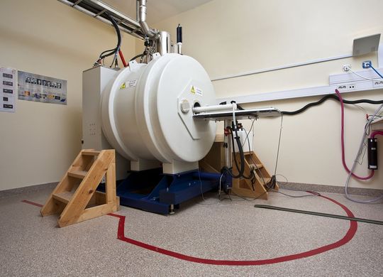
Bruker Pharmascan 70/16
A 7.0 Tesla small animal magnetic resonance (MR) scanner was installed at the Vivarium, University of Bergen, in December 2004 and was upgraded with new hardware in December 2012.
This MR scanner can be used for non-invasive diagnostic studies on small laboratory animals (rats, mice, small fish).
Pathological and physiological processes can now be followed in a precise manner in the animal body by utilizing a set of MR imaging techniques combining visualisation of anatomy, diffusion, perfusion, and MR spectroscopy.
The MR machine is specifically suitable for performing longitudinal studies of disease development and treatment efficacy.
Announcements and Events
Lab Equipment
Technical details about the MRI scanner, operating software, animal monitoring equipment etc.
People
Getting Started
Check out this page if you are planning on doing MRI experiments. Remember that you need to receive training before you can do experiments on your own, so please plan ahead.
How To
This page has a lot of information on the operation of the 7T scanner, scanning protocols and pulse sequences, data transfer and analysis, animal physiology and anaesthesia, training, safety, how to register as a user, etc.
Booking the MRI
Takes you directly to MIC's booking website.
Policies and Regulations
Safety and Operator Training
Contains links to lecture material from an in-depth course in small animal MRI.
Forms
Gallery
Under construction.
Research
Information about some of the research groups who have used the MR scanner in the past.


