Frits Thorsen´s group
Group members
Frits Thorsen, group leader
Terje Sundstrøm, PhD Student
A novel animal model for characterization and treatment of melanoma metastasis
Background
Brain metastasis is a complication occurring in one third of all cancer patients. The annual incidence in the US is 170 000. Brain metastases is the commonest intracranial cancer, appearing 10 times more often than primary brain tumors (1). The most common sources of metastatic brain lesions involve the lung (50-60%), breast (15-20%), skin (5-10%) and gastrointestinal tract (4-6%).
Patients with melanoma metastatic spread to the central nervous system (CNS) have poor prognosis (2, 3). Treatment strategies available today are ineffective, with low response rates, as current chemotherapeutic agents are ineffective. Clearly there is a need for suitable animal models in which spontaneous CNS metastases develop when cancer cells metastasize from the primary lesion in a manner that reflects the patient setting. We have currently developed such models, which will enable us to study the biology and the treatment of melanoma brain metastases.
Development of new metastatic animal models
We have recently developed a novel model system where tumor cells from human melanoma brain metastases are injected systemically in immunodeficient mice and metastasize to the mouse brain (4). This animal model recapitulates most steps of the metastatic cascade, and a better understanding of the molecular mechanisms behind tumor metastasis may thus be obtained.


- Development of metastatic cell lines from a melanoma brain metastasis
An important question is whether melanoma cells excised from brain metastases in patients would metastasize selectively to the brain in a mouse model system, if the tumor cells are injected into the bloodstream.
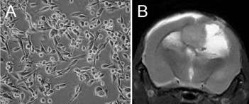
In order to study this we developed several tumor cell lines from a brain metastases harvested from a patient with malignant melanoma (Figure 1A). To verify the tumorigenicity of the cell line, melanoma cells were injected directly into the brains of immunodeficient nod/SCID mice. During a time period of 3-4 weeks, large lesions developed (Figure 1B).
- Insertion of reporter genes to study tumor cell colonization and growth in vivo.
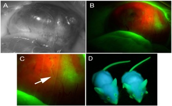
We then generated new cell lines, by labeling the tumor cells with suitable reporter genes. First, we inserted the DsRed reporter gene into the H1 cell line, using a lentiviral vector, resulting in the H1_dsRed cell line. This cell line was embedded in matrigel, and injected subcutaneously into eGFP nod/SCID mice (5). After around 4 weeks, large tumors developed subcutaneously (Figure 2).
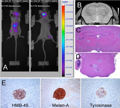
Systemic in vivo tumor development can also be detected by bioluminescence imaging of the animals. Thus we used a retroviral GFP/Luciferase construct, and obtained a 100% GFP/Luciferase positive cell line after FACS sorting. These cells were injected into the left ventricle of the heart in nod/SCID mice, and tumor development was studied by bioluminescence imaging. After 3-8 weeks, the animals developed new tumors in most organs of the mouse body (Figure 3A). T1w/T2w MRI showed multiple metastases within the CNS (Figure 3B). Around 30 micrometastases (sizes 0.1-0.8mm) were detected in the animal brain by histology (Figure 3C, 3D). The melanocytic properties of the tumors were confirmed by immunohistochemistry (Figure 3E).
Ongoing research activities
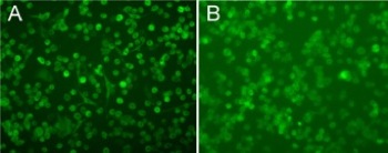
We have already injected the H1_GFP_Luc metastatic melanoma cell line into several mice, and collected tumors from lung, bone, CNS and ovary. These tumors have been plated, and new cell lines have been established (Figure 4). We are currently reinjecting these new cell lines into mice, to see if the tumor cells home specifically to the organ they were established from.
The H1_GFP_Luc tumor cells are also being injected into the blood stream of immunodeficient mice, and the colonization and growth of new tumors are being detected using advanced preclinical MR imaging. It has previously been shown that breast cancer cells prelabelled with micron-sized iron oxide particles (MPIOs) and therafter injected intra-cardially into mice, can be tracked by MRI as single cells in the mouse brain (6). In a similar manner, we are labeling the H1_GFP_Luc melanoma cell line with Fedex (7), and performing T2 and T2* weighted MR imaging.
Brain metastases are very often highly vascularized, a process driven primarily by VEGF. In this part of the project, we are specifically targeting the neovascularization process using bevacizumab treatment. Bevacizumab, which is a VEGF antibody, has previously been used in advanced, primary cancers (8-10). Just recently, the antibody has also been FDA approved for use on patients with brain metastasis. However, there is limited knowledge on treatment effects and biological responses after bevacizumab treatment on metastatic brain tumors. Our metastatic brain tumor modell develops highly angiogenic tumors, and thus mimic the vascular, metastatic brain tumors seen in patients. After injecting the H1_GFP_Luc cell line in the left ventricle of the mice, we are treating the animals with clinically relevant bevacizumab doses, injected intraperitoneally once a week for 3 weeks. The animals are then be studied with MR imaging.
See also Terje Sundstrøm PhD outline at https://wikihost.uib.no/mriwiki/index.php/Terje_Sundstrøm_PhD_project
References
- Santarelli JG, Sarkissian V, Hou LC, Veeravagu A, Tse V. Molecular events of brain metastases. Neurosurg Focus 22(3):E1, 2007.
- Tarhini AA, Agarwala SS. Management of brain metastasis in patients with melanoma. Curr Opin Oncol 16:161-166, 2004).
- JuanYin J, Tracy K, Zhang L, Munasinghe J, Shapiro E, Koretsky A, Kelly K. Noninvasive imaging of the functional effects of anti-VEGF therapy on tumor cell extravasation and regional blood volume in an experimental brain metastasis model. Clin Exp Metastasis 26:403-414, 2008.
- Wang J, Daphu I, Pedersen PH, Miletic H, Hovland R, Mørk S, Bjerkvig R, Tiron C, McCormack E, Micklem D, Lorens JB, Immervoll H, Thorsen F. A novel brain metastases model developed in immunodeficient rats closely mimics the growth of metastatic brain tumours in patients. Neuropathol Appl Neurobiol 2010 (Resubmitted).
- Niclou S, Danzeisen C, Eikesdal HP, Wiig H, Brons NH, Poli AM, Svendsen A, Torsvik A, Enger PØ, Terzis AJ, Bjerkvig R. A novel EGFP-expressing immunodeficient mouse model to study tumor-host interactions. FASEB J 22:3120-3128, 2008.
- Heyn C, Ronald JA, Ramadan SS, Snir JA, Barry AM, MacKenzie LT, Mikulis DJ, Palmieri D, Bronder JL, Steeg PS, Yoneda DJ, MacDonald IC, Chambers AF, Rutt BK, Foster PJ. In vivo MRI of cancer cell fate at the single-cell level in a mouse model of breast cancer metastasis to the brain. Magn Res Med 56:1001-1010, 2006.
- Babic M, Horak D, Trchova M, Jendelova P, Glogarova K, Lesny P, Herenyk V, Hajek M, Sykova E. Poly(L-lysine)-modified iron oxide particles for stem cell labelling. Bioconjugate Chem 19:740-750, 2008.
- Perez DG, Suman VJ, Fitch TR, Amatruda T 3rd, Morton RF, Jilani SZ, Constantinou CL, Markovic SN. Phase 2 trial of carboplatin, weekly paclitaxel, and biweekly bevacizumab in patients with unresectable stage IV melanoma: a North Central Cancer Treatment Group study, N047A. Cancer 115:119-127, 2009.
- Gonzalez-Cao M, Viteri S, Diaz-Lagares A, Gonzaléz A, Redondo P, Nieto Y, Espinos J, Chopitea A, Ponz M, Martín-Algerra S. Preliminary results of the combination of bevacizumab and weekly Paclitaxel in advanced melanoma. Oncology 74:12-16, 2008.
- Scicher N, Paulitschke V, Swoboda A, Kunstfeld R, Loewe R, Pilarski P, Pehamberger H, Hoeller C. Erlotinib and bevacizumab have synergistic activity against melanoma. Clin Cancer Res 15:3495-3502, 2009.

